What is a thyroid scan?
Review:
A thyroid scan (thyroid scintigraphy) is a diagnostic nuclear medicine test that provides information about the structure and function of the thyroid. The nuclear medicine physician is able to see the size, shape, function, and position of the thyroid gland.
Your will swallow a capsule or liquid containing a radiopharmaceutical called Iodine-123. Iodine-123 has a tiny amount of radioactive molecules in it.
A special camera, called a gamma camera, is used to take pictures of the thyroid at four and 24 hours after the radiopharmaceutical has been ingested.
When might a thyroid scan be needed?
A thyroid scan can help assess:
- neck masses
- hypothyroidism
- hyperthyroidism
- ectopic thyroid
- thyroid malignancy
- thyroglossal duct cyst
- benign diffuse goiter
- thyroiditis
- radiation therapy planning
How the Test is Performed
The test is done in this way:
- You are given a pill that contains a tiny amount of radioactive iodine. After swallowing it, you wait as the iodine collects in your thyroid.
- The first scan is usually done 4 to 6 hours after you take the iodine pill. Another scan is usually done 24 hours later. During the scan, you lie on your back on a movable table. Your neck and chest are positioned under the scanner. You must lie still so that the scanner gets a clear image.
- The scanner detects the location and intensity of the rays given off by the radioactive material. A computer displays images of the thyroid gland. Other scans use a substance called technetium instead of radioactive iodine.
.jpg)

Risks
All radiation has possible side effects. The amount of radioactivity is very small, and there have been no documented side effects. Women who are pregnant or breastfeeding should not have this test.
The radioactive iodine leaves your body through your urine. You should not need to take special precautions, such as flushing twice after urinating, for 24 to 48 hours after the test because the dose of radioactive iodine is very low. Ask your provider or the radiology/nuclear medicine team performing the scan about taking precautions.
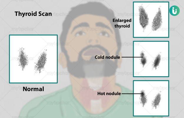
What Abnormal Results Mean
- A thyroid that is enlarged or pushed off to one side could be a sign of a tumor.
- Nodules absorb more or less iodine and this will make them look darker or lighter on the scan. A nodule is usually lighter if it has not taken up the iodine (often called a 'cold' nodule). If part of the thyroid appears lighter, it could be a thyroid problem. Nodules that are darker have taken up more iodine (often called a 'hot' nodule). They can be overactive and may be the cause of an overactive thyroid.
- The computer will also show the percentage of iodine that has collected in your thyroid gland (radioiodine uptake). If your gland collects too much iodine, it may be due to an overactive thyroid. If your gland collects too little iodine, it may be due to inflammation or other damage to the thyroid.

Ablasi thyroid